Home › Unlabelled › Foot Muscles Mri Anatomy / Ankle MRI: Torn #Achilles tendon in 78 yo woman who fell ...
Foot Muscles Mri Anatomy / Ankle MRI: Torn #Achilles tendon in 78 yo woman who fell ...
Foot Muscles Mri Anatomy / Ankle MRI: Torn #Achilles tendon in 78 yo woman who fell .... There are 10 intrinsic muscles located in the sole of the foot. The muscles acting on the foot can be divided into two distinct groups; If more detail is needed, however, an orthopedic doctor will likely want to do magnetic resonance imaging (mri). This chapter will address the specific anatomy and physiology relevant to mri. Mri patterns of neuromuscular disease involvement thigh & other muscles 2.
The foot is a part of vertebrate anatomy which serves the purpose of supporting the animal's weight and allowing for locomotion on land. Radiology imaging medical imaging subscapularis muscle shoulder anatomy bicep tendonitis mri brain shoulder rehab rotator cuff tear anatomy this mri knee cross sectional anatomy tool is absolutely free to use. The muscles acting on the foot can be divided into two distinct groups; If more detail is needed, however, an orthopedic doctor will likely want to do magnetic resonance imaging (mri). Parts of the brain & function.
Mri Lower Extremity Anatomy - Human Anatomy from www.imaios.com 2, tensor fasciae latae m. Mri of the ankle and feet. They act collectively to stabilise the arches of the foot, and individually to control movement of the digits. The movement and stability of the arch is controlled by intrinsic and extrinsic muscles. Foot muscles dorsal region & #8211; 550 x 385 jpeg 31 кб. In addition to the three above mentioned muscles there are more structures lying in the medial group of the plantar fascia e.g. 3, vastus medialis & intermedius muscles.
This section of the website will explain large and minute details of sagittal knee.
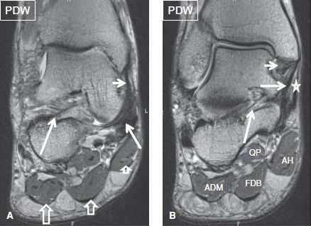
In addition to the three above mentioned muscles there are more structures lying in the medial group of the plantar fascia e.g. Simplified radiological anatomy of the foot. Related posts of foot muscle anatomy mri. This chapter will address the specific anatomy and physiology relevant to mri. 2 muscle layers superficial layer: Foot muscles are grouped into 4 regions: They are individual positioned medial to their respective tendon of the flexor digitorum longus. However, the intrinsic muscles are largely ignored by. Mri imaging of the foot • examinations are usually divided into : Near normal foot mri for reference. Mri of the ankle and feet. With increasing mri scanning resolution, future studies may be able to investigate the volumes of individual intrinsic foot muscles. 3, vastus medialis & intermedius muscles.
Foot muscles dorsal region & #8211; The muscles that control the movements of the foot originate in the lower leg and are attached the bones in the foot with tendons. 850 x 882 png 268 кб. The foot is a complex structure with many articulations and multiple degrees of freedom that play an important role in static posture and dynamic activities. Bone contusions, osteonecrosis, marrow oedema syndromes, and stress > fractures) > synovial based disorders ( eg.
The Ankle | Musculoskeletal Key from musculoskeletalkey.com Indications for foot mri scan. The muscles acting on the foot can be divided into two distinct groups; 3, vastus medialis & intermedius muscles. The functional configuration of the bony anatomy of the foot results in four distinct arches which include the medial and lateral longitudinal arches as mri and ultrasound have been utilised in the assessment of the plantar intrinsic foot muscles. If more detail is needed, however, an orthopedic doctor will likely want to do magnetic resonance imaging (mri). They mostly work together to support the arches of the foot. Parts of the brain & function. 2 muscle layers superficial layer:
Magnetic resonance imaging (mri) is the method of choice for detecting soft tissue structure and abnormalities 58, 59.
Muscle anatomy model 12 photos of the muscle anatomy model anatomy muscle models arm, muscle anatomy model, muscle anatomy model amazon, muscle anatomy model labeled, shoulder muscle anatomy model, human muscles. Intrinsic muscle atrophy and toe deformity in the diabetic. The anatomy of the foot and common foot problems. You can divide the foot muscles into groups based on their locations — either in the sole of the foot (the bottom or plantar area) or the dorsum they don't work as intricately as the small muscles in the hands; Foot muscles dorsal region & #8211; The tendon of the flexor hallucis. Simplified radiological anatomy of the foot. Legs come in all shapes and sizes, ranging from portly and stout, to the artists usually begin their study of the legs by focusing on the athletic type, because the shapes of the muscles are more easily seen. Magnetic resonance imaging (mri) image showing foot. They act collectively to stabilise the arches of the foot, and individually to control movement of the digits. Related posts of foot muscle anatomy mri. Tibialis anterior, extensor hallucis longus, extensor digitorum longus. 850 x 882 png 268 кб.
The muscles that control the movements of the foot originate in the lower leg and are attached the bones in the foot with tendons. Legs come in all shapes and sizes, ranging from portly and stout, to the artists usually begin their study of the legs by focusing on the athletic type, because the shapes of the muscles are more easily seen. Composite video showing multiple mri images including: Mri imaging of the foot • examinations are usually divided into : The foot is a part of vertebrate anatomy which serves the purpose of supporting the animal's weight and allowing for locomotion on land.
The Blog Life of Lady Blackwood: Football Follies from 1.bp.blogspot.com Feet and ankles ankle muscle anatomy of foot muscles of foot muscles foot foot muscles anatomy muscle drawing foot ligaments anatomy of the foot. Intrinsic muscle atrophy and toe deformity in the diabetic. Ankle and hind foot examination. This page provides a gallery of images that presents the anatomical structures found on thigh mri. Learn anatomy faster and remember everything you learn. The foot is a part of vertebrate anatomy which serves the purpose of supporting the animal's weight and allowing for locomotion on land. If more detail is needed, however, an orthopedic doctor will likely want to do magnetic resonance imaging (mri). Mri imaging of the foot • examinations are usually divided into :
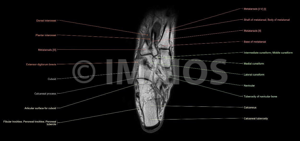
A magnetic resonance imaging (mri) was performed on a cross section of the foot with anatomical structures labeled as arteries, muscles.
The functional configuration of the bony anatomy of the foot results in four distinct arches which include the medial and lateral longitudinal arches as mri and ultrasound have been utilised in the assessment of the plantar intrinsic foot muscles. Foot muscles are grouped into 4 regions: Together, the upper and lower legs and the feet make up half the length of the human figure. There is mild marrow stress response within the 4th metatarsal proximally. Learn anatomy faster and remember everything you learn. Tibialis anterior, extensor hallucis longus, extensor digitorum longus. Mri of the ankle and feet. This article discusses anatomy, supply and function of the muscles found on the medial plantar aspect/ sole of the foot. The foot is a complex structure with many articulations and multiple degrees of freedom that play an important role in static posture and dynamic activities. Like the fingers, the toes have flexor and extensor muscles that power their movement and play a large role in balance. 850 x 882 png 268 кб. Magnetic resonance imaging (mri) image showing foot. Magnetic resonance imaging (mri) is the method of choice for detecting soft tissue structure and abnormalities 58, 59.
This section of the website will explain large and minute details of sagittal knee foot muscles mri. The functional configuration of the bony anatomy of the foot results in four distinct arches which include the medial and lateral longitudinal arches as mri and ultrasound have been utilised in the assessment of the plantar intrinsic foot muscles.

comment 0 comments
more_vert